Health Sciences
Şaban Tiryaki
August 2019
Series Editor: Vesile Şenol
Referees: Kadirhan Doğan, Eşref Kızılkaya
Redactor: Berk İlke Dündar
Cover Design: Nazile Arda Çakır
Page Design: Evren Demiryürek
Tiryaki, Şaban.
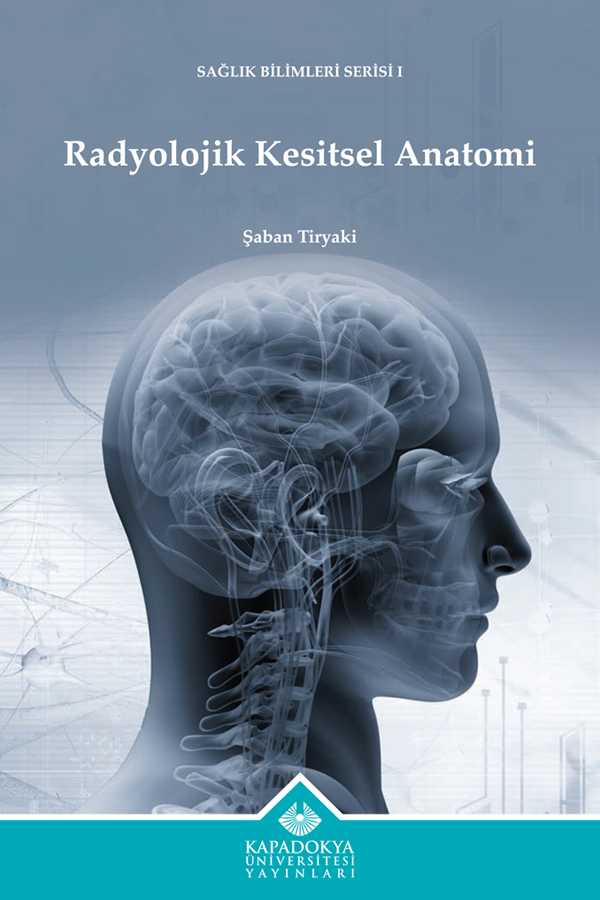
Radiological Cross-Sectional Anatomy
287 pages, 195×270 mm.
There are references.
This book has been prepared to follow the curriculum of the Cross-Sectional Radiological Anatomy course taught especially in the associate-degree Medical Imaging Techniques program. The book presents cross-sectional views of the anatomy of the human body, CT images, and MR image orientation in the sagittal, axial and coronal planes in an easily understandable way. The book strives to reinforce learning with multiple choice tests at the end of each lecture, and makes various references. The normal anatomy depicted in the study can serve as a reference when interpreting pathologies for those working in the fields of neurology, neurosurgery, orthopedics, physiotherapy and rehabilitation, and those with an interest in cross-sectional radiological anatomy, and can aid both students specializing in Radiology and practicing Radiologists. The book is oriented toward Medical Imaging Techniques program students and those with an interest in this field.

Şaban Tiryaki graduated from the Faculty of Medicine of Uludağ University in 1990. After working as a physician in the central health clinic in Üzümlü, Erzincan, and thereafter in Kaman, Kırşehir, he passed the examination for specialty in medicine and went on to study in the Radiation Oncology Department of Ankara Numune Hospital in 1993. He completed his specialization in Radiology in the Faculty of Medicine of Cumhuriyet University and became a Radiology Specialist in 1998. He has worked as a Radiology Specialist in Erzincan State Hospital, Ürgüp State Hospital, Nevşehir State Hospital, Private Versa Hospital and Private Aşıkpaşa Hospital. He has since undergone training in second level obstetric ultrasonography. In his working life, he has performed dual contrast gastrointestinal examinations, DSA, MRI, CT, USG, Doppler USG, 4D USG, trucut breast biopsy and percutaneous drainage. His specialty thesis compared the success of venography with colour Doppler ultrasonography for the diagnosis of deep vein thrombosis. Tiryaki is currently lecturing in the Medical Imaging Techniques program of Cappadocia University.
In 2000, he published an article on his thesis topic in the journal of the Faculty of Medicine of Cumhuriyet University. In 2007, he published a case report on Dysgerminoma arising in Swyer Syndrome in The Internet Journal of Pathology with colleagues from Erzincan State Hospital. The author’s book Radiological Cross-Sectional Anatomy was published by Cappadocia University Publications in 2019.
Click to access the book.